Bright signal around cells in a photo easily makes people think "the peptide went in." But signal may also remain on the surface, come from free dye, or be affected by handling. First clarify what the image can answer, then discuss entry into cells.

Fluorescence tracks labeled material
Fluorescent labels help us observe, but they also change molecular structure. Label position, linker, and dye properties may affect binding, distribution, and fluorescence behavior. Richard et al.'s classic cell-penetrating peptide research reminds us that factors such as fixation and surface binding affect understanding of uptake mechanisms. This is background from other cell-penetrating peptide studies and cannot be directly used as uptake results for a given LL-37 batch. [1]
First distinguish the outer cell edge from the cell interior
Ordinary two-dimensional images superimpose information from different depths, so signal on or above the cell surface may appear to fall within the cell outline. Combining confocal sections, membrane markers, or other suitable methods helps further determine location. But every method has resolution and processing limits, and one cannot rely only on a single selected representative photo.
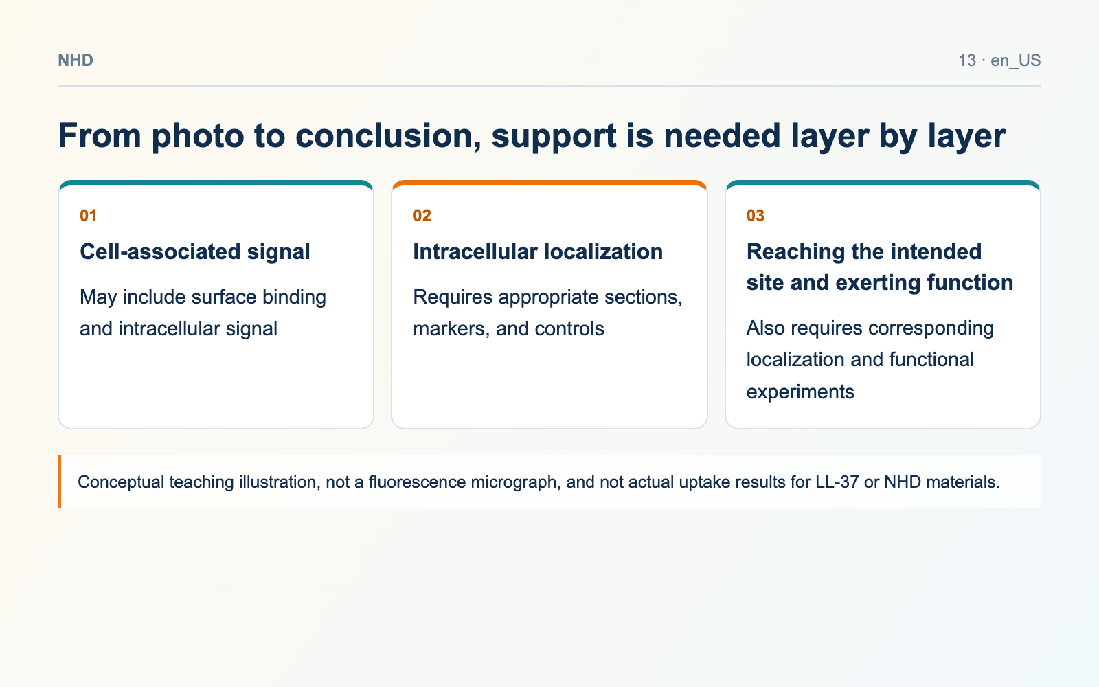
From photo to conclusion, support is needed layer by layer
- Cell-associated signal: May include surface binding and intracellular signal
- Intracellular localization: Requires appropriate sections, markers, and controls
- Reaching the intended site and exerting function: Also requires corresponding localization and functional experiments
Controls determine which interpretations still hold
Depending on the experimental goal, one can set up unlabeled cells, dye alone, labeled material, and necessary unlabeled material controls, and check the washing process. If considering surface signal quenching, removal, or other discrimination methods, first confirm their effects on cells and readouts. Weaker signal after some treatment does not automatically mean all previous signal was outside the cell; the treatment itself may also change what is observed.
Brightness is not a direct scale of molecule number
Dye amount, illumination, exposure, pH, local environment, and signal quenching all affect brightness. Imagine two teaching images: one with longer exposure and another with shorter exposure. Even with the same sample, they will look different. Therefore, comparing brightness requires consistent, suitable acquisition and processing rules, and original images should be retained. When quantification is needed, the normalization method and measurement range should also be stated.
Entering the cell does not mean reaching the site of action
Intracellular signal may be located in regions such as endocytic vesicles and does not automatically mean the molecule has reached the cytoplasm, nucleus, or intended site of action. If the research aims to answer delivery or functional questions, more specific localization and functional evidence is needed. Writing "associated with cells," "entered cells," and "produced a specific effect" as three different conclusions can reduce leaps in image interpretation.
When purchasing, match the label to the experiment
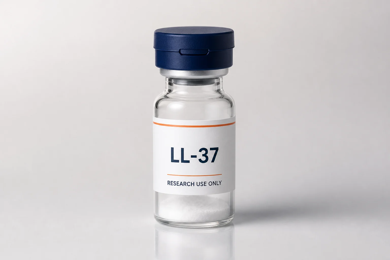
If a fluorescently labeled form of LL-37 is needed, specify the sequence, labeling site, dye, and linker, and check them against the instrument channel. Also confirm what identity and quality information exist for the ordinary material and the labeled material separately. The catalog LL-37 card is for understanding related research materials; the specific labeled version must be confirmed separately, and one must not treat the ordinary peptide as a validated fluorescent probe just because the product name is the same.
Original sources
Check the study conditions and material records separately. Calculations labeled as examples are not batch test results.
 LL-37