When the blank area in a culture dish narrows, it means cell coverage has changed. But the cells may have moved into the space, increased in number, or both. When reading peptide repair studies, this distinction matters.

First, see how the assay creates a blank area
In the classic scratch assay, a gap is created in a cultured cell layer, and then researchers observe how cells cover that area over a set period. The methods paper by Liang et al. describes this approach to studying migration. The blank area is not a human wound, and closure in a culture dish does not mean tissue repair has been completed. Cell type, culture conditions, initial density, and imaging time can all affect what is observed. [1]
Calculate the closure percentage from area
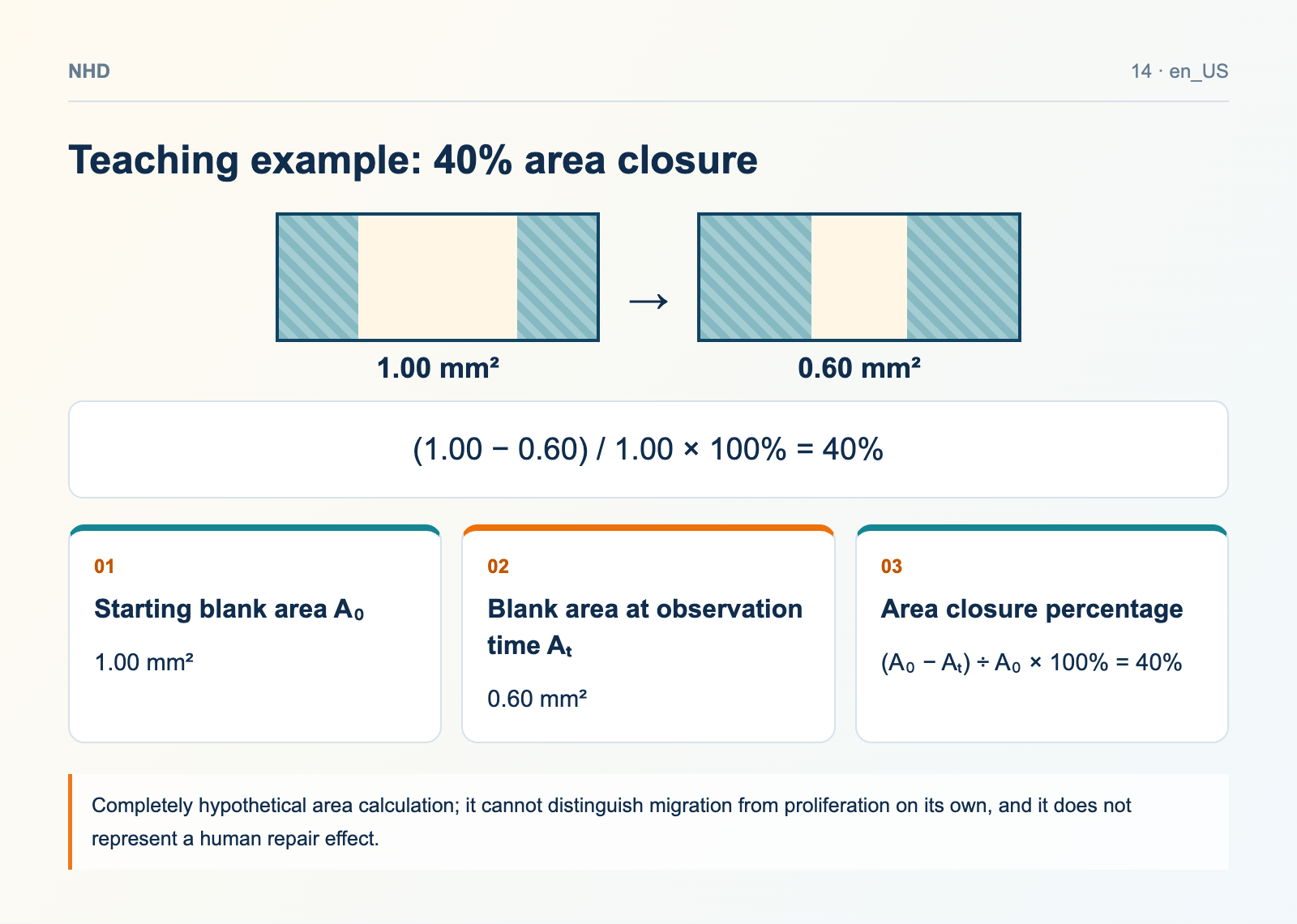
A teaching example: the starting blank area is 1.00 mm², and at a given time point 0.60 mm² remains, so the area closure percentage = (1.00−0.60) ÷ 1.00 × 100% = 40%. If another group starts with an area of 0.80 mm², you cannot directly compare the final remaining areas of the two groups. The starting state and normalization rules need to be consistent. The data in this example are entirely hypothetical and do not represent migration speed, cell number, or any real effect of any peptide.
Teaching example: 40% area closure
- Starting blank area A₀: 1.00 mm²
- Blank area at observation time Aₜ: 0.60 mm²
- Area closure percentage: (A₀ − Aₜ) ÷ A₀ × 100% = 40%
Both movement and proliferation make the blank smaller
Cells moving along the surface can cover the gap; cell division increasing numbers can also change coverage. To discuss the contribution of migration, you need to combine an appropriate observation time, an assessment of proliferation, and necessary controls. If an anti-proliferative treatment is used, you also need to assess whether it affects cell viability and migration itself. You cannot assume that other effects have completely disappeared just because some treatment was applied.
Before comparing photos, check comparability
Are the initial gaps similar? Were the same cells, culture medium, solvent, and observation intervals used? Were images taken from pre-specified positions? Are the area segmentation and boundary rules consistent? If you pick only the fields with the best closure, the results may deviate from the performance of the whole group. Keeping the original images, field numbers, and analysis rules helps others verify the work, rather than leaving only the final composite.
Multiple photos do not necessarily equal multiple independent samples
Taking ten photos of the same culture well mainly adds observational information about that well; it does not equal ten independent experiments. Study design needs to distinguish fields, culture wells, independent preparations, or cell sources, and statistical analysis should match the hierarchy. When describing results, state clearly what was measured and what the unit of comparison was; "more closure" cannot be expanded on its own into "promotes human wound healing."
How to relate GHK-Cu and BPC-157 materials
These materials may appear in various repair-related studies, but the cells, treatments, and results in each paper need to be checked individually. When purchasing, first confirm the actual molecular form and batch documentation; when running experiments, keep records of preparation, controls, and observations. Product cards serve as an entry point for further verification; do not turn a result from cultured cells into a human efficacy claim for that product.
Original sources
Check the study conditions and material records separately. Calculations labeled as examples are not batch test results.
 BPC-157
 GHK-Cu