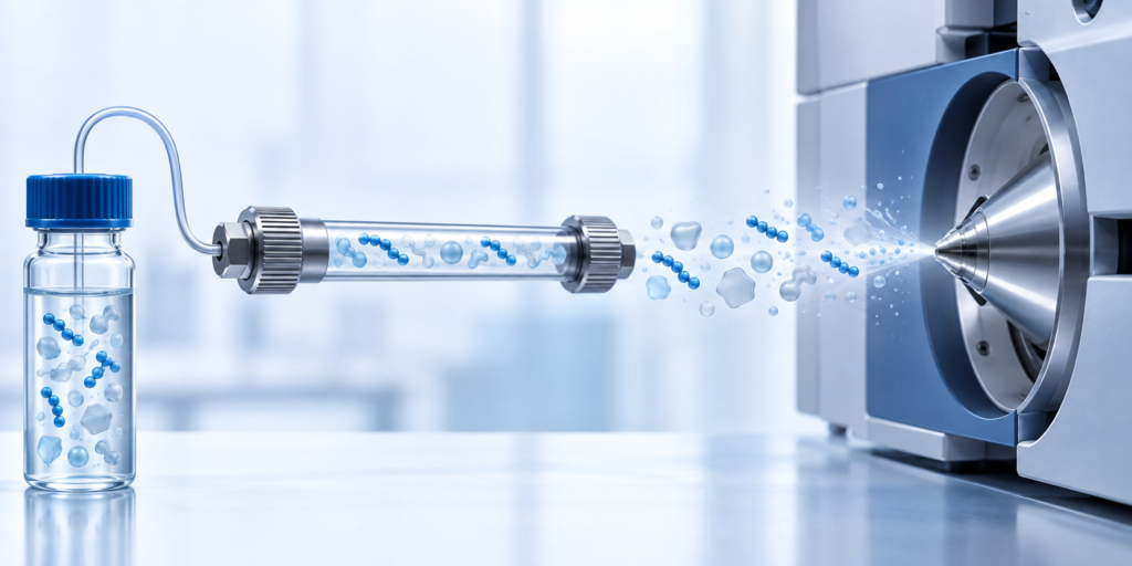
An intense peptide signal in neat solvent can fade in plasma without a loss of peptide mass. Coeluting matrix components can change electrospray response. A calibrator prepared only in neat solution may therefore misrepresent the response in a biological sample. The practical decision is to test candidate surrogate peptides in the intended matrix and select a quantifier on validated behavior, rather than signal alone.
A measured example from rituximab analysis
In a study comparing LC–MS/HRMS and LC–MS/MS methods for rituximab in human plasma, investigators generated surrogate peptides after protein preparation and digestion. Their Table 7 reports matrix effects for several candidate peptides. For the HRMS method, six plasma sources were spiked at 50 µg/mL rituximab. Ion suppression differed markedly: FSGS averaged −67% (range −54% to −78%), pQVQ averaged −11% (reported range +15% to −35%), and pQIVL averaged −93% (range −88% to −95%). The authors used pQIVL only as a qualifier because its matrix effect was large in both MS methods. [1, §2.3.3, Table 7]
| HRMS surrogate peptide | Mean reported matrix effect | Tested setting |
|---|---|---|
| FSGS | −67% | Six human plasma sources; rituximab spike 50 µg/mL |
| pQVQ | −11% | Same study and HRMS setting |
| pQIVL | −93% | Same study and HRMS setting; used as qualifier |
The LC–MS/MS arm used four plasma sources, spiked at 15 and 300 µg/mL. Its Table 7 reports −17% mean suppression for pQVQ and −22% for pQIVL. Do not merge those numbers with the HRMS values into one generic “peptide matrix effect”: the instruments, source counts and spike levels differ. [1]
A practical evaluation design
Choose more than one candidate peptide where the protein sequence permits. Confirm each candidate’s identity and separation, then compare response in neat solution, post-extraction matrix and fully processed matrix. The contrast between neat and post-extraction response helps identify ionization effects; the contrast between post-extraction and processed response can reveal preparation loss. Check multiple independent matrix lots and the concentration range that the assay must measure. Track the internal-standard-normalized matrix factor if a suitable isotope-labeled standard is available. The paper used full-length labeled rituximab as an internal standard, which can follow more preparation steps than a peptide added only after digestion. The timing of standard addition must match what the assay claims to correct.
Treat matrix effects as analyte- and method-specific. A less suppressed peptide still needs acceptable selectivity, calibration, precision and digestion behavior. A highly suppressed candidate can sometimes serve as a qualifier if it remains reliable for identity confirmation, as in the reported pQIVL example; that does not make it a sound sole quantifier. In the same paper, the authors reported calibration and precision results for their two validated methods, but these do not prove transferability to another antibody or instrument. [1, §§2.3.2–2.3.3]
Decision record for a surrogate peptide
For each candidate, record: (a) sequence uniqueness and fragment-ion support, (b) digestion yield and missed-cleavage forms, (c) matrix-factor distribution across independent matrix lots at low and high concentrations, (d) retention-time separation from interference, and (e) performance after internal-standard normalization. Keep rejected candidates and the reason for rejection. An apparent high neat-solvent response cannot substitute for these checks.
The ICH M10 guideline independently defines matrix effect as changed analyte response due to matrix components. For regulatory bioanalytical validation it calls for low and high QC replicates across at least six matrix sources/lots, with justified flexibility for rare matrices. That is a relevant design benchmark, not an automatic requirement for every exploratory research assay; the paper’s HRMS experiment also used six plasmas, while its MS/MS arm used four. [2, §§1.3 and 3.2.3]
Boundary and next step
The measured values come from rituximab surrogate peptides in human plasma, not free peptide standards or NHD products. They show why a sequence’s analytical response cannot be assumed from another sequence. For interpretation of a specific peptide’s exact mass and fragmentation, use the LC–MS identity guide; for questions about the meaning of chromatographic area and content, see the purity-versus-assay guide. Neither page replaces matrix-matched validation.
Primary source and claim trail
- Development, Validation, and Comparison of Two Mass Spectrometry Methods (LC-MS/HRMS and LC-MS/MS) for the Quantification of Rituximab in Human Plasma. Molecules. 2021;26:1383. DOI 10.3390/molecules26051383, PMID 33806585. Locations: §2.3.3 and Table 7 (plasma source counts, spike levels, peptide-specific matrix effects and pQIVL qualifier decision); §2.3.2 (calibration context); Methods (full-length stable-isotope rituximab). Table values were checked in the primary full-text XML.
- International Council for Harmonisation. ICH M10: Bioanalytical Method Validation and Study Sample Analysis, final guideline, 2022. Locations: §§1.3 and 3.2.3; regulatory scope and independent matrix-lot design. This is authoritative method guidance, not evidence of the study’s measured suppression values.
Sources & editorial method
AI-assisted editorial draft; independent Codex source, image and seven-language review; human professional review not claimed
2 linked records are listed in the references below. Read the editorial and AI-assistance policy.
How to interpret this article
This article summarizes third-party records and does not establish the identity, quality, safety or efficacy of any catalog lot.
Research-use boundary: Catalog materials discussed on this website are for laboratory research, development and manufacturing use only, not for human or veterinary use. This content is not medical advice and does not provide administration instructions.
Primary records and authoritative sources
- Development, Validation, and Comparison of Two Mass Spectrometry Methods (LC-MS/HRMS and LC-MS/MS) for the Quantification of Rituximab in Human PlasmaSource 1. §2.3.3 and Table 7 (plasma source counts, spike levels, peptide-specific matrix effects and pQIVL qualifier decision); §2.3.2 (calibration context); Methods (full-length stable-isotope rituximab). Table values were check
- International Council for Harmonisation. ICH M10: Bioanalytical Method Validation and Study Sample Analysis, final guideline, 2022. Locations: §§1.3 and 3.2.3; regulatory scope and independent matrix-lot design. This isSource 2. §§1.3 and 3.2.3; regulatory scope and independent matrix-lot design. This is authoritative method guidance, not evidence of the study's measured suppression values.