Candida albicans is a fungus, not a bacterium. Researchers used live-cell imaging to watch what happens after LL-37 contacts it, wanting to know where the peptide goes first and what changes occur in the cell. When reading images like these, the key is to separate "seeing a signal" from "proving an effect."

First, know what you are observing
Candida albicans has a cell wall and a cell membrane, and can grow in different forms. The cell wall is not another name for the cell membrane; their locations and compositions differ. Observing that a fluorescent peptide stays around the cell may suggest contact with outer structures, but a plain two-dimensional image alone does not necessarily tell you precisely whether it is on the wall, on the membrane, or already inside the cell. Studies usually need to combine more specific labels and assays.
Live-cell imaging mostly shows the order of events
A study published in 2014 compared the interactions of peptides such as LL-37 and CATH-2 with Candida albicans, combining live-cell imaging with other observation methods. Fluorescent labeling helps researchers track the peptide's location; compared with looking only at endpoint photos, continuous observation can show the sequence of events. However, something happening first in time does not automatically mean it is the sole cause, and the fluorescent label itself may also affect molecular behavior. [1]
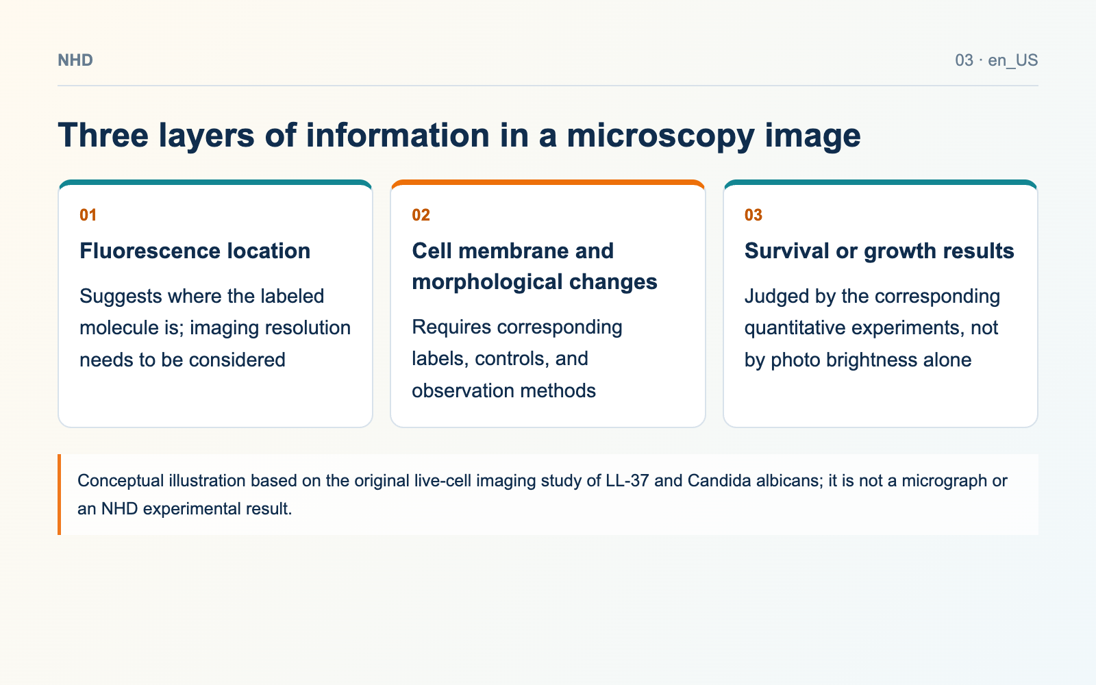
Three layers of information in a microscopy image
- Fluorescence location: Suggests where the labeled molecule is; imaging resolution needs to be considered
- Cell membrane and morphological changes: Requires corresponding labels, controls, and observation methods
- Survival or growth results: Judged by the corresponding quantitative experiments, not by photo brightness alone
Localization, membrane changes, and survival are best read separately
The paper observed that LL-37 rapidly associates with the Candida albicans cell membrane. This localization result should be understood together with membrane permeability, morphological changes, and antifungal assays. Imagine a teaching example: the signal around the cell is very bright, but the survival indicator changes very little; in that case, what you can say first is "binding or aggregation occurred," and you cannot skip the other experiments and declare that the fungus was killed. This example only illustrates how to interpret results; it does not correspond to real experimental values. [1]
When comparing with bacterial experiments, don't just match concentrations
When the same concentration of LL-37 is used against two microorganisms, the results can still be affected by culture medium, salt, cell number, growth state, and observation time. The outer structures of fungi and bacteria also differ. When comparing papers, check the strain, experimental conditions, and assay endpoints first, rather than comparing only a single minimum concentration. Cells that look more "broken" in micrographs also cannot directly replace comparable quantitative results.
To read an image, you can follow this order
First find the microorganism and peptide names in the legend, then see what each dye labels. Next check the scale bar, time points, exposure method, and whether untreated, unlabeled, and dye-only controls are included. Finally, see which independent experiments the authors used to support their interpretation. If brightness needs to be compared, exposure and image-processing rules must be consistent; some images are only suitable for showing location, not for using pixel intensity to calculate molecule numbers.
When preparing similar experiments, specify materials and labels first
When inquiring about LL-37 or fluorescent labeling services, you should state the sequence, label type, label position, linker, and the excitation and emission ranges the instrument can use. Plain LL-37 and labeled LL-37 need to be identified separately and cannot be glossed over with the same abbreviation. The original paper describes the samples it used and cannot prove that NHD catalog batches have the same results; product information and experimental records still need to be checked one by one.
Original sources
Check the study conditions and material records separately. Calculations labeled as examples are not batch test results.
 LL-37