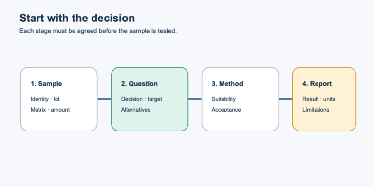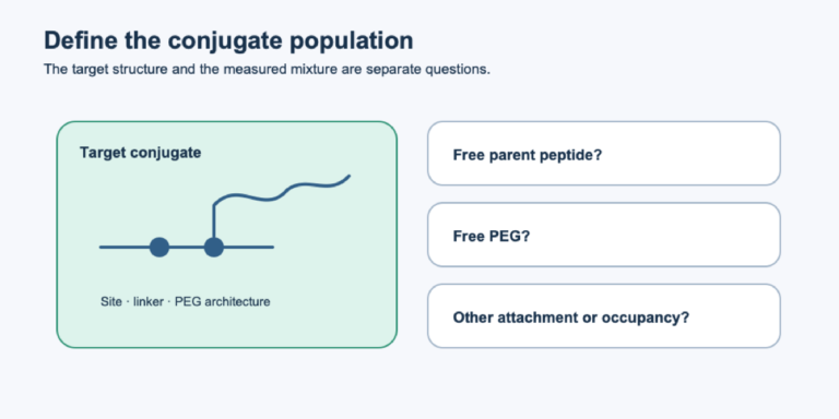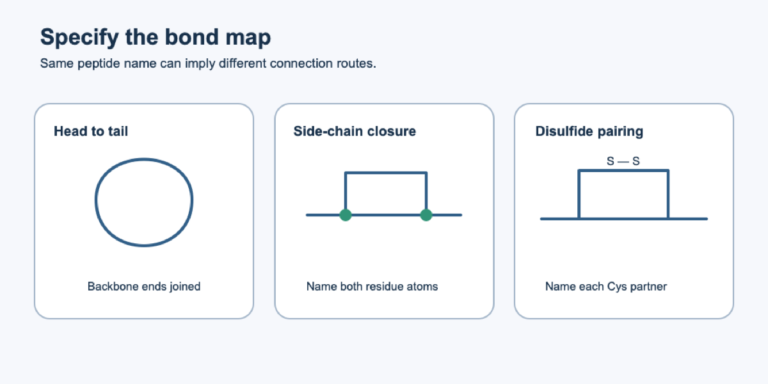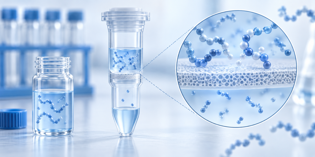
A 30 kDa filter does not mean every peptide below 30 kDa passes into the filtrate. Molecular-weight cutoff (MWCO) describes a membrane’s separation behavior under stated conditions; the measured result also depends on peptide binding to carrier proteins, membrane interaction, sample pretreatment and the downstream detection method. The question for a laboratory is not “Which cutoff is smallest?” but “Which preparation retains the peptide information that this assay needs?”
What the primary experiment actually compared
Hölttä and colleagues developed a method to recover endogenous peptides from human cerebrospinal fluid (CSF) for offline LC–MALDI mass spectrometry. They evaluated 10, 30 and 50 kDa ultrafiltration devices for peptides in an approximately 700–5,000 Da analysis window. Their paper reports that 10 kDa devices retained many peptides of interest, while 50 kDa devices allowed appreciable albumin through; the authors selected 30 kDa for that CSF workflow. The 10- and 50-kDa observations were reported without quantitative comparative data, so they cannot support a numerical ranking for other samples. [1, Results: “Sample preparation”]
The authors then tested sample pretreatment before the 30 kDa step. In aliquots from a pooled CSF sample, 2,445 detected compounds appeared without pretreatment and 3,543 after 20% acetonitrile (ACN) pretreatment. Editorial calculation from those two reported counts: 1,098 additional detected features, about 45% more relative to the untreated condition. Those are instrument-detected features, not peptide recovery percentages or newly validated biomarkers. At 40% ACN, detected features decreased; the authors proposed co-precipitation as one possible explanation. Their final workflow yielded 3,000–4,000 peptide-like features and identified 730 peptides by MS/MS, illustrating that feature detection and confident identity are separate counts. [1, Figure 1 and Abstract]
| Study condition | Reported result | What it measures |
|---|---|---|
| Pooled CSF, no pretreatment before filtration | 2,445 detected compounds | Features observed by the study’s LC–MALDI method |
| Pooled CSF, 20% ACN before filtration | 3,543 detected compounds | Features observed by the same method |
| Final CSF peptidomics workflow | 730 MS/MS-identified peptides | Sequence-level identifications, not a filter recovery rate |
The paper also mentions approximately 76% recovery for a 1,350 Da peptide, but explicitly attributes that figure to filter product documentation, not its own validation. It should not be presented as the study’s measured recovery. [1, Results: “Sample preparation”]
How to turn the observation into a method check
For a defined target peptide, measure its signal or amount in the starting sample, filtrate, membrane retentate and any rinse, using a validated calibration strategy. Include a matrix-matched control that bypasses the filter when feasible. This distinguishes a filter-associated deficit from low signal caused downstream by ion suppression. Repeat with the intended sample volume and protein load; a result from pooled CSF need not transfer to plasma, cell medium or a purified standard.
If the goal is discovery peptidomics, track identified sequences and missingness as well as total feature count. More features after pretreatment might reflect release from carrier proteins or improved ionization, rather than a simple increase in peptide mass recovered. If the goal is a targeted assay, assess each analyte’s recovery and reproducibility. No single MWCO or organic-solvent percentage should be copied from this paper without checking protein precipitation, chemical stability, assay compatibility and local safety controls.
An independent HLA immunopeptidomics comparison identified another loss pathway: peptides could remain associated with the ultrafiltration membrane, so its workflow collected an acetonitrile-based filter rinse and analyzed it with the filtrate. That does not prove the same rinse is suitable for CSF, but it makes a filter-retained fraction worth measuring before concluding that a peptide was absent from the starting sample. [2]
Four records to release a filtration result
- Record membrane chemistry, MWCO, centrifugation conditions, starting volume and protein load.
- Report target signal or sequence IDs in input, filtrate, retentate and any validated rinse. State when a fraction could not be measured.
- Include a bypass or post-filtration spike to check whether changed matrix composition altered MS response rather than recovery.
- Report missing peptide identities separately from total detected features; neither count alone is a mass balance.
Evidence limit and next step
This is a laboratory analytical-method study using CSF. It provides a useful example of why membrane rating, sample pretreatment and downstream identification must be assessed together. It does not establish a general CSF diagnostic claim, the performance of any NHD consumable, or the quality of any peptide lot. Pair filter-recovery checks with the LC–MS identity guide when a target’s identity matters, and document preparation conditions alongside reported results.
Primary source and claim trail
- Hölttä M, et al. Peptidome Analysis of Cerebrospinal Fluid by LC-MALDI MS. PLOS ONE. 2012;7:e42555. DOI 10.1371/journal.pone.0042555; full text PMC3412831, PMID 22880031. Locations: Abstract (3,000–4,000 features; 730 identities), Results and Discussion → “Sample preparation” and Figure 1 (10/30/50 kDa qualitative comparison; 2,445/3,543 features; ACN concentration). The 76% is identified there as manufacturer documentation and is excluded from our measured-results table.
- Mild Acid Elution and MHC Immunoaffinity Chromatography Reveal Similar Albeit Not Identical Profiles of the HLA Class I Immunopeptidome. Original comparative immunopeptidomics study; its ultrafiltration methods discuss peptide retention and an acetonitrile-based filter rinse. Use: an independent surface-retention mechanism, not a CSF recovery value or a validated rinse recipe for this article’s matrix.
Sources & editorial method
AI-assisted editorial draft; independent Codex source, image and seven-language review; human professional review not claimed
2 linked records are listed in the references below. Read the editorial and AI-assistance policy.
How to interpret this article
This article summarizes third-party records and does not establish the identity, quality, safety or efficacy of any catalog lot.
Research-use boundary: Catalog materials discussed on this website are for laboratory research, development and manufacturing use only, not for human or veterinary use. This content is not medical advice and does not provide administration instructions.
Primary records and authoritative sources
- Hölttä M, et al. Peptidome Analysis of Cerebrospinal Fluid by LC-MALDI MS. PLOS ONE. 2012;7:e42555.Source 1. Abstract (3,000–4,000 features; 730 identities), Results and Discussion → “Sample preparation” and Figure 1 (10/30/50 kDa qualitative comparison; 2,445/3,543 features; ACN concentration). The 76% is identified there as m
- Mild Acid Elution and MHC Immunoaffinity Chromatography Reveal Similar Albeit Not Identical Profiles of the HLA Class I ImmunopeptidomeSource 2. See primary-source and claim-trail entry in article.md