


A fluorescent peptide can be correctly labelled and still produce a disappointing signal if the instrument cannot excite or detect that label well. Before choosing a colour, check the instrument’s light source and filters.
A fluorescent label has two useful wavelength numbers
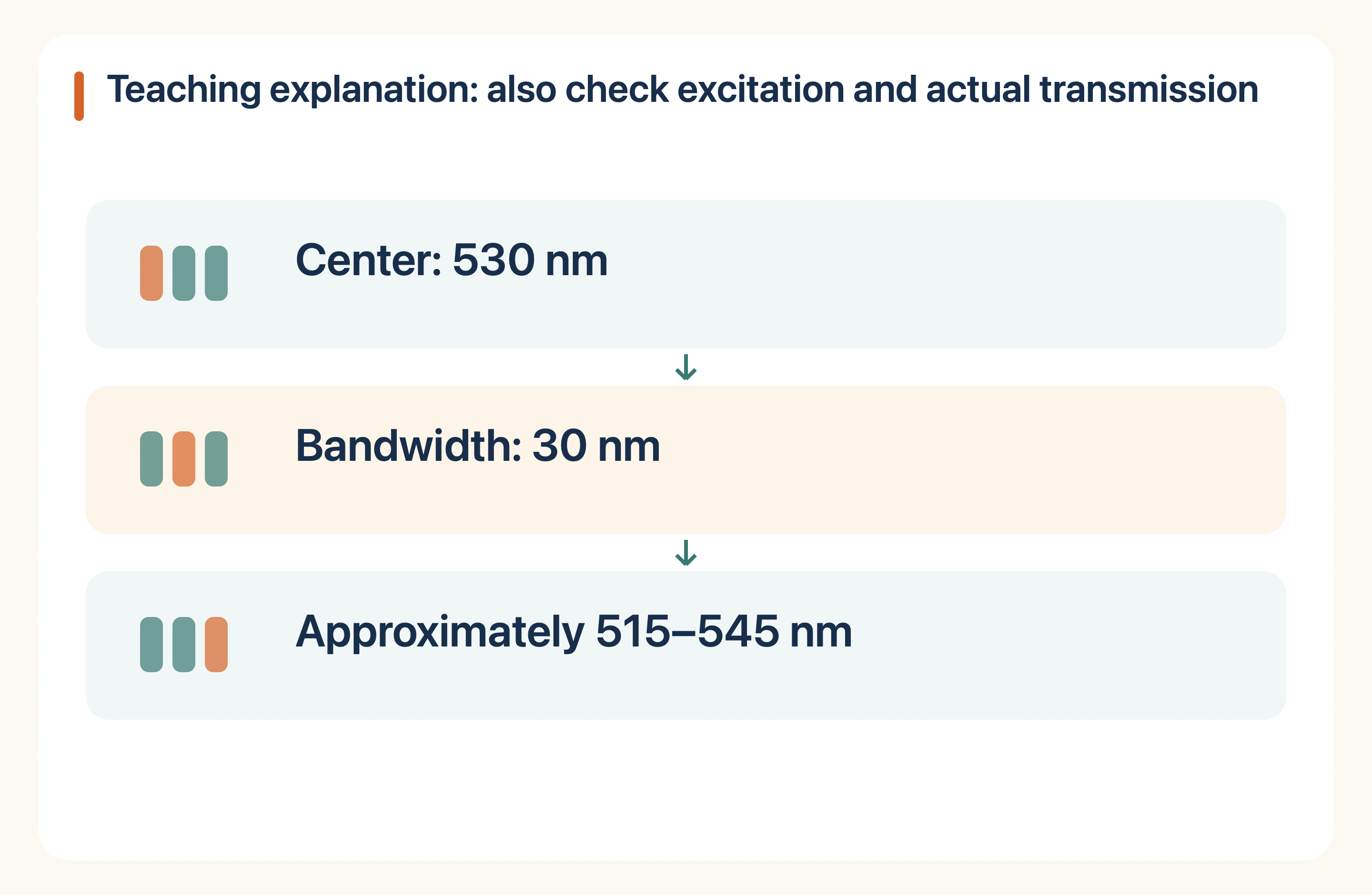
Excitation describes the light used to stimulate the dye. Emission describes the light it gives back. Thermo Fisher lists Alexa Fluor 488 with excitation/emission maxima of 494/519 nm and Alexa Fluor 647 with maxima of 650/668 nm. These are manufacturer dye-reference values, not measurements from an NHD peptide batch. The surrounding chemistry and measurement conditions can affect the spectrum.
Colour names are only a starting point
“Green” or “red” does not specify a complete optical setup. A detector also needs an appropriate emission window, and a light source needs to excite the dye sufficiently. Peak wavelengths are not hard cut-offs: dyes have broader spectra. Compare the actual instrument configuration with the dye information rather than matching a colour word alone.
The label can change the material
Attaching a dye adds a chemical group and changes the expected mass. The attachment position and any linker belong in the order specification. A labelled peptide should not be assumed to behave exactly like the unlabelled peptide in every assay. If comparison matters, plan an unlabelled control or another suitable control with the research team.
What to send with a quotation request
Give the sequence, preferred dye, attachment site, linker if required, and instrument model or excitation/emission settings. Ask which labelled forms are feasible and what purity and identity evidence is included. Also ask how free dye or unlabelled peptide is evaluated when relevant. Agree on the material and analytical scope before treating a generic “fluorescent peptide” quotation as complete.
Next step with NHD
The related product cards below identify catalogue peptide families. They do not confirm availability of biotinylated, fluorescent, isotope-labelled or library versions. For a modified material, specify the exact design in a custom inquiry.
Start with the Labelled custom peptides. For a quotation, send NHD your sequence or product name, quantity and required documents. Confirm the exact form, feasibility, available testing and delivery terms before ordering.
Sources
Public references explain the topic; they are not NHD batch evidence. Research materials are for laboratory research, development and manufacturing, not human or veterinary use.
Sources & editorial method
AI-assisted writing, source checks and illustrated cover.
1 linked records are listed in the references below. Read the editorial and AI-assistance policy.
How to interpret this article
Public data and labeled teaching calculations; not NHD batch results. Confirm project and document availability.
Research-use boundary: Catalog materials discussed on this website are for laboratory research, development and manufacturing use only, not for human or veterinary use. This content is not medical advice and does not provide administration instructions.
Primary records and authoritative sources
- Using the Fluorescence SpectraViewerUsing the Fluorescence SpectraViewer
Continue with structured records
Move from this editorial analysis to the product identity, filtered evidence and controlled terminology behind it.



 KPV
 LL-37